User guide
Examples
Here are some examples...
Download structure, select, align, and write
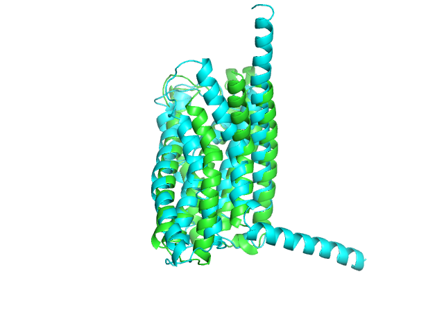
Download a pdb, select desired atoms, align the structures, and save the structures to pdb files.
from protutils.pdb import PDBFile


# Download 4k5Y.pdb from the RCSB Protein Data Bank
pdb = PDBFile.fetch('4K5Y')

# Select residues less than 263 on chain A, then chain B
chain_a = pdb.select(chain__eq='A', nres__lt=263)
chain_b = pdb.select(chain__eq='B', nres__lt=263)

# Align selection with another selection that contains the same number of atoms
aligned_chain_b = chain_b.align(chain_a)
# prints: RMSD = 0.983960869568

# write structures to pdb files
chain_a.write_pdb('chain_a.pdb')
chain_b.write_pdb('chain_b.pdb')
aligned_chain_b.write_pdb('aligned_chain_b.pdb')
Now, we can visualize the structures in pymol.
load chain_a.pdb
load chain_b.pdb
load aligned_chain_b.pdb

zoom
show cartoon
ray
png example.png
This produces the following image
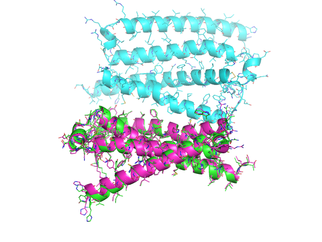
The align method works selections of equal size (e.g. different conformers of the same molecules). It will fail for molecules with different number of atoms. To align different molecules, use the cealign method.
from protutils.pdb import PDBFile


pdb = PDBFile.fetch('4K5Y')
chain_a = pdb.select(chain='A')  # equivalent to pdb.select(chain__eq='A')
chain_b = pdb.select(chain='B')

# compare the number of atoms in the selection
len(chain_a) == len(chain_b)
# returns False

# the align method would fail for these selections
aligned_chain_b = chain_b.cealign(chain_a)
# prints RMSD = 0.874903919378  (RMSD of alpha carbons)

# write structures
chain_a.write_pdb('chain_a.pdb')
chain_b.write_pdb('chain_b.pdb')
aligned_chain_b.write_pdb('aligned_chain_b.pdb')
Visualizing the saved structures gives
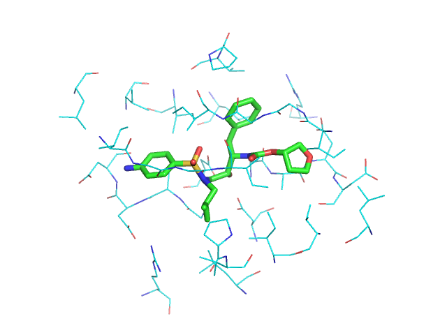
Select binding site residues
Selection helper functions allow quick selections of protein, ligand, backbone, and side chain atoms. Distance selections between two objects can be made. Most methods return a new object, allowing chaining methods together.
from protutils.pdb import PDBFile


# write a small helper function
def residues_for_selection(selected, pdbfile):
    """Get all residues atoms for a selection
    """
    residues = {atm.nres for atm in selected}
    return pdbfile.select(nres__isin=residues)


pdb = PDBFile.fetch('1HPV')

# Select ligand
ligand = pdb.ligand()

# select protein atoms with 5 Angstroms of the ligand
atoms = pdb.protein().within(5.0, ligand)

prot = residues_for_selection(atoms, pdb)

ligand.write_pdb('ligand.pdb')
prot.write_pdb('prot.pdb')
Visualizing gives
Structure analysis: Ramachandran plot
Create a Ramachandran plot of a protein.
from protutils.pdb import PDBFile


pdb = PDBFile.fetch('4K5Y')
protein = pdb.protein()  # remove HETATM records
protein.ramachandran_plot()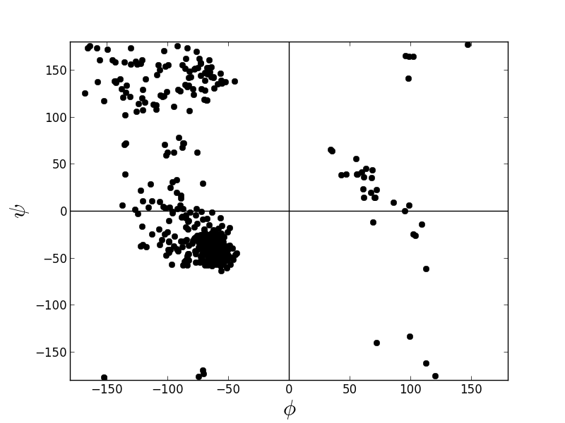
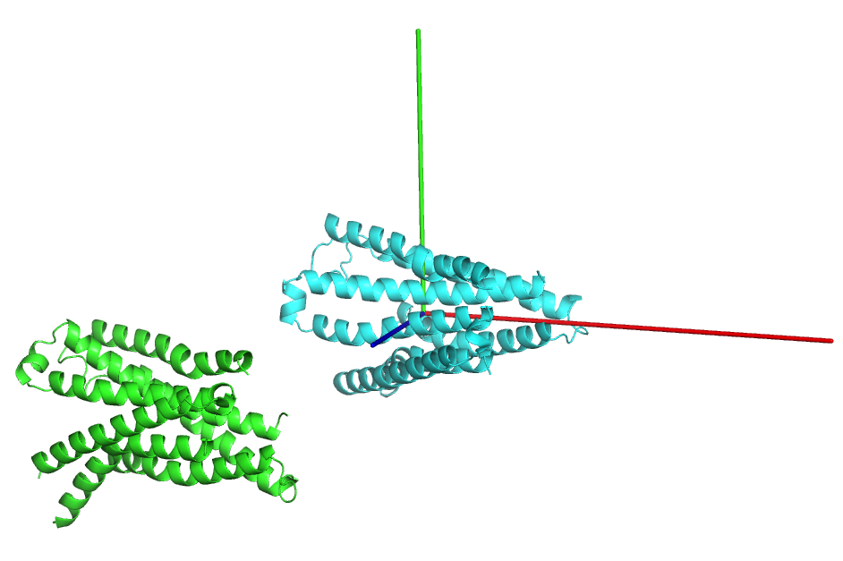
Orient principle axes of a protein along Cartesian axes
It is sometimes useful to orient structures such that the centroid lies at the origin and the principle axes of the protein align with the x, y, and z axes.
from protutils.pdb import PDBFile

pdb = PDBFile.fetch('4K5Y')
protein = pdb.select(chain='A', nres__lt=1000).protein()

orient = protein.orient()

protein.write_pdb('4K5YA.pdb')
orient.write_pdb('4K5YA_0.pdb')
Visulaize the transformed structure in pymol (Cartesian axes indicated by rgb lines).
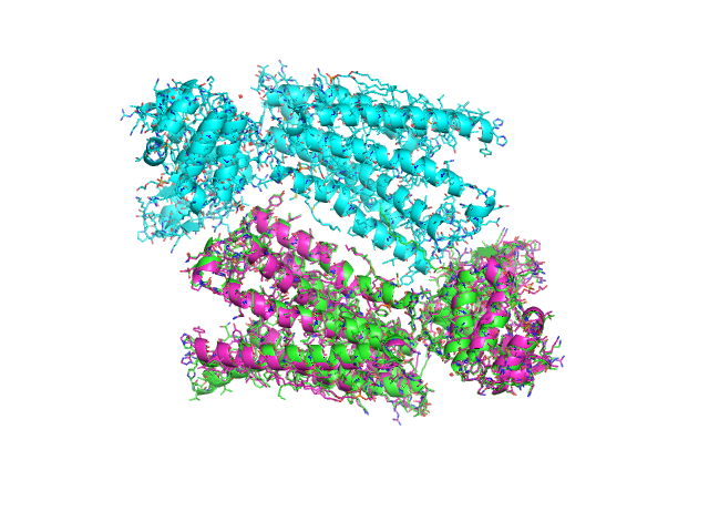
Search for similar Structures
Find similar proteins using PDB BLAST.
from protutils.pdb import PDBFile
from protutils.ncbi.blastp import BLASTPDBRecord

pdb = PDBFile.fetch('4K5Y')
chain_a = pdb.select(chain='A', nres__lt=1000)
# Get sequence
sequence = chain_a.sequence.replace('-', '')

# search for similar sequences to 4K5Y_A
query = BLASTPDBRecord(sequence)

# Get top hit
PDB = query.get_best()['pdb']
similar = PDBFile.fetch(PDB)
similar.select(chain='A', nres__lt=1000)

# Compare aligned structures
aligned = similar.cealign(chain_a)

chain_a.write_pdb('4k5y_A.mod.pdb')
aligned.write_pdb('{0}_A.mod.pdb'.format(PDB))
Now, compare the structures: